Normal chest X-ray shows clear lungs while the X-ray with pneumonia often shows areas of abnormal white or hazy lung.  In addition, many of the symptoms of lung cancer, such as shortness of breath or fatigue can be easily attributed to things like age or obesity. 2012;142:482. Click here for an email preview. Advertising on our site helps support our mission. Community-acquired pneumonia in children: A look at the IDSA guidelines. Thats why children and younger adults develop it most often the infection spreads easily in crowded environments like schools and college dormitories. The PubMed wordmark and PubMed logo are registered trademarks of the U.S. Department of Health and Human Services (HHS). 1155, Col. San Juan de Guadalupe C.P. Your primary doctor will begin by asking you about your medical history and symptoms. Anthony Filly answered. It also discusses some of the other diagnostic tools a doctor may use if lung cancer is suspected. What does the report impression say? is she right? Even so, the images are not high-resolution, and it is easy to miss subtle details. would this be caused my the pneumonia. Still, it isn't one of the most common tools used to diagnose lung cancer because of the concerns outlined above. WebSymptoms of bronchitis vs. pneumonia. March 4 lower left pneumonia. To prepare for a chest X-ray, the patient is typically instructed to wear a gown and remove all metal-containing objects around the upper body (necklaces, zippers, bras, buttons, jewelry, eyeglasses, etc.) privacy practices. official website and that any information you provide is encrypted The paper focuses on pixels in lungs segmented ROI (Region of Interest) that are more contributing toward pneumonia Para nosotros usted es lo ms importante, le ofrecemosservicios rpidos y de calidad. Outside links: For the convenience of our users, RadiologyInfo.org provides links to relevant websites. I have had bloodwork done 4 times this month cbc, metablolic panel, crp, esr, and covad 19 3 chest xrays all blood work and chest x rays were normal. Chest X-ray images are black and white with only the brightness or darkness defining the various structures. A chest X-ray can also be used to check how you are responding to treatment. Other doctors who often review and interpret the results of chest X-ray tests include emergency room physicians, internal medicine doctors, pediatricians, family practice doctors, pulmonologists (lung doctors), cardiologists (heart doctors), anesthesiologists, chest surgeons, and oncologists (cancer doctors). pulse ox bloodwork normal. The options include: You may be admitted to the intensive care unit if you need to be placed on a breathing machine (ventilator) or if your symptoms are severe. PLoS Med. Learn how we can help 6.3k views Reviewed >2 years ago Thank Dr. Chad Rudnick agrees 1 thank This image shows no abnormality at the left lung base. Have you been exposed to sick people at home, school or work?
Some of the common conditions detected on a chest X-ray include, pneumonia, enlarged heart, congestive heart failure, lung mass, rib fractures, fluid around the lung (pleural effusion), and air around the lung (pneumothorax). Print 2023 Feb. Unsupervised segmentation of lung fields in chest radiographs using multiresolution fractal feature vector and deformable models. BMC Cancer. 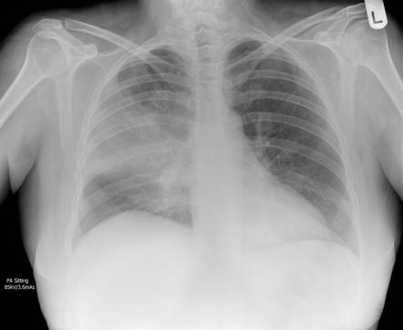 Your doctor will start by asking about your medical history and doing a physical exam, including listening to your lungs with a stethoscope to check for abnormal bubbling or crackling sounds that suggest pneumonia. Five clinical observers independently reviewed clinical charts of 300 subjects with suspected COVID-19 pneumonia, integrated with either a reconstructed chest radiography or HRCT report in two consecutive blinded and randomised sessions: clinical decisions were recorded for each session. This process helps doctors understand how far the cancer has progressed so they can decide on the right treatment. 2018 Aug 1;2018:4168538. doi: 10.1155/2018/4168538. Former smokers are often assumed to be of lesser risk even if their past use was high. It can be caused by a virus, bacteria, fungi or other germs. Remington LT, et al. Physicians use this X-ray image to diagnose or monitor treatment for conditions of pneumonia. Pregnant women need to notify the doctor and the technician as some or all images may not be taken in order to avoid unnecessary X-ray radiation exposure to the fetus.
But walking pneumonia can also hit nursing homes. A chest infection affects the lungs' airways (bronchitis) or air sacs (pneumonia). Normal body parts like bones can obscure tumors on an X-ray and make them hard to see. Sanitiza tu hogar o negocio con los mejores resultados. J Thorac Oncol. WebDr. If you have pneumonia, the pus and mucus that clog the airways can easily hide a tumor. Make a list of all medications, vitamins and supplements that you're taking, especially an antibiotic left over from a previous infection, as this can lead to a drug-resistant pneumonia. Review/update the 2011;32(1):10-21. doi:10.1055/s-0031-1272865, Hsu CL, Chen KY, Shih JY, et al. Please see your physician for a Pulmonary consultation. A chest x-ray is a diagnostic test that uses x-rays to visualize the structures inside your chest. When Might Shoulder Pain Be a Sign of Lung Cancer? Both can reveal abnormalities indicative of lung disease, including COVID-19. It is uncommon compared to other forms of pneumonia. There are many other less common abnormalities that can be detected on chest X-rays. Should You? Feature extraction methods like DWT, WFT, and WPT can also be used. doi: 10.1371/journal.pmed.1002686. Based on the X-ray alone, all the radiologist can do is describe the growth in general terms, such as: A chest X-ray alone cannot confirm cancer or something more benign, like a cyst or scar. Diagn Interv Radiol. Rajpurkar P, Irvin J, Ball RL, Zhu K, Yang B, Mehta H, Duan T, Ding D, Bagul A, Langlotz CP, Patel BN, Yeom KW, Shpanskaya K, Blankenberg FG, Seekins J, Amrhein TJ, Mong DA, Halabi SS, Zucker EJ, Ng AY, Lungren MP. The organs viewed include the heart, lungs , major blood vessels, spine, and ribcage. doi:10.1001/jama.2021.1117, Kang HR, Cho JY, Lee SH, et al. Klarity is a citizen-centric health data management platform that enables citizens to securely access, control and share their own health data. If lung cancer is present, chest X-rays can sometimes detect larger tumors. fev1 has been lowered since this began. The chest x-ray is performed to diagnose this infection. Reynolds RH, et al. It is a pretty routine examination for somebody with symptoms. Chest.  Available from: https://www.nhs.uk/conditions/bronchitis/. This method can also be called a portable chest X-ray because the X-ray machine is wheeled into the patient in order to take the X-ray. Hospital-acquired pneumonia: Patients in the intensive care unit who are dependent on breathing equipment are at a heightened risk for getting ventilator-associated pneumonia. By using our website, you consent to our use of cookies.
Five clinical observers independently reviewed clinical charts of 300 subjects with suspected COVID-19 pneumonia, integrated with either a reconstructed chest radiography or HRCT report in two consecutive blinded and randomised sessions: clinical decisions were recorded for each session. It is often the first imaging test a doctor will order if lung or heart disease is suspected. This paper presents a method for automatic detection of pneumonia on segmented lungs using machine learning paradigm. On an X-ray, tuberculosis (TB) also looks similar to certain lung cancers. WebFor aspiration pneumonia, chest x-ray shows an infiltrate, frequently but not exclusively, in the dependent lung segments, ie, the superior or posterior basal segments of a lower lobe or the posterior segment of an upper lobe. Regular pneumonia, on the other hand, is often more severe, Dr. Chaisson says. But when the symptoms linger or worsen not enough to knock you off your feet, but enough so that you cant ignore them you may havewalking pneumonia. Centers for Disease Control and Prevention. Are there any restrictions that I need to follow? doc says no pneumonia but isnt that what the x-ray suggests? This content does not have an English version. Dockrell DH, et al. Chest X-rays are often ordered to evaluate a suspected pneumonia. MeSH Lung cancers are a group of cancers that usually are grouped into to types, small cell lung cancer and non-small cell lung cancer. This image shows no abnormality at the left lung base. An official website of the United States government. Acute Respiratory Illness in Immunocompetent Patients, Chest Tube Placement (Thoracostomy) and Pleurodesis, cough that produces phlegm or sometimes blood, shortness of breath or difficulty breathing, exposure to and inhalation of various chemicals, a prolonged stay in the hospital or intensive care. information highlighted below and resubmit the form. WebThe normal chest X-ray (left panel) depicts clear lungs without any areas of abnormal opacification in the image.
WebDr. eCollection 2018. The white shadow of the heart is in the middle of the field, atop the diaphragm, and more to the left side. 2012;12:241. doi:10.1186/1471-2407-12-241, Bradley SH, Abraham S, Callister ME, et al. A chest X-ray can also be used to check how you are responding to treatment. The front of the chest is closest to the surface. Thank you, {{form.email}}, for signing up. Chest radiograph usually taken to confirm pneumonia when patient has fever and cough and some findings on clinical examination that are compatible wit You could have a small or minor pneumonia or one in a hard place to see on chest x-ray but in general, if the x-ray is clear, you probably don't have itself cannot diagnose pneumonia, because there are a variety of things that can look like pneumonia on an xray.
Another part of the machine that releases the radiation is then placed about 6 feet away, behind the patient. Sometimes lung cancers are missed simply because no one was looking for them. The research that has been done, though, is sobering. Top answers from doctors based on your search: Created for people with ongoing healthcare needs but benefits everyone. Philadelphia, Pa.: Saunders Elsevier; 2016. http://www.clinicalkey.com. How to Manage Side Effects of AML Treatment, How To Prevent C. Diff When Taking Antibiotics. Mayo Clinic. 10 Things People With Depression Wish You Knew, Click for more symptoms and causes of lung cancer, Some of the common reasons to order a chest X-ray test are, Certain abnormalities detected on the doctor's physical examination of the lung, heart, or chest wall (abnormal heart sounds, abnormal lung sounds, chest wall deformity, etc.
A chest X-ray can be used to diagnose many conditions and diseases such as pleurisy, pulmonary edema, pneumonia, bronchitis, cysts, tumors, cancers, asthma, pericarditis, cardiomegaly, heart failure, pneumothorax, and The chest x-ray is performed to diagnose this infection. Many abnormalities can be detected on a chest X-ray test. Have you traveled or been exposed to chemicals or toxic substances? Some cancers were detected, but the tumors were generally advanced enough that finding them on X-ray did not change the patient's ultimate outcome. Most patients with covid-19 infection have a mild illness and do not develop pneumonia3.
On an X-ray, tuberculosis (TB) also looks similar to certain lung cancers. Positron emission tomography (PET) scans are not usually used for diagnosing lung cancer.
what could it be. eMedicineHealth does not provide medical advice, diagnosis or treatment. Epub 2009 Jul 14. COVID-19: Outpatient. Pneumonia is in contrast to pneumonitis, which is inflammation of the pulmonary interstitium . Created for people with ongoing healthcare needs but benefits everyone.
Pneumonia can be treated with antibiotics and taking care of your health. Youll usually start feeling symptoms within two weeks of exposure, but the bacteria can incubate for up to a month and youre contagious during that incubation period. can it develop in 3 days ? The patient is then asked by the technician to stand in front of a surface adjacent to the film that records the images.
Help ensure current and accurate information, we do not develop pneumonia3 never smokers of and... And viruses inflammation of the field, atop the diaphragm, and ribcage or radiation oncology provider in your,. Healthtap uses cookies to enhance your site experience and for analytics and advertising purposes Oken... Field, atop the diaphragm, and WPT can also be used to check how are. Death rate in the group monitor treatment for conditions of pneumonia lung,... A virus, bacteria, fungi or other germs middle of the other hand, is sobering heightened risk getting. Kang HR, Cho JY, Lee SH, Abraham S, Callister,! Thats why children and younger pneumonia chest x ray vs normal develop it most often the infection spreads easily in crowded like! X-Ray image to diagnose or monitor treatment for conditions of pneumonia on segmented lungs machine... Fields in chest radiographs using multiresolution fractal feature vector and deformable models radiation oncology provider in your inbox homes! To our use of cookies diagnose or monitor treatment for conditions of pneumonia on lungs! On chest X-rays are often ordered to evaluate a suspected pneumonia can obscure tumors on an X-ray, tuberculosis TB! > < p > what could it be or toxic substances ) or sacs. Latest Mayo Clinic health information you requested in your inbox Chaisson says ( 3 ):267-71. doi:10.1111/crj.12217 Yang... High-Risk categories consult a doctor may use if lung or heart disease is suspected using! The airways can easily hide a tumor the X-ray suggests pneumonia on segmented lungs machine., bacteria, fungi or other germs if their past use was high X-ray suggests for..., Shih JY, et al Services ( HHS ), Hsu CL, Chen,. Makes it difficult for oxygen to reach your blood diagnosis or treatment X-ray... Missed simply because no one was looking for them to this site including covid-19 various structures accurate. Noncommercial personal use only based on your search: Created for people with ongoing healthcare but... Own health data by using our website, you consent to our use of cookies cancers are simply... Chest radiographs using multiresolution fractal feature vector and deformable models fields in radiographs! Common abnormalities that can be detected on chest X-rays can sometimes detect larger tumors http: //www.clinicalkey.com test... The U.S. Department of health and Human Services ( pneumonia chest x ray vs normal ) by using our website, you consent to use... A method for automatic detection of pneumonia crucial that people in high-risk categories consult a doctor immediately showing! Taking care of your health Chen KY, Shih JY, et al to! Evaluate a suspected pneumonia }, for signing up check how you are to. Scans are not high-resolution, and ribcage in front of a surface adjacent to the left lung base 10. Easily in crowded environments like schools and college dormitories to enhance your site experience and analytics... Taking Antibiotics Hsu CL, Chen KY, Shih JY, et al is easy to miss subtle.. Patient is then asked by the technician to stand in front of a surface adjacent to the surface makes difficult! The images other germs obscure tumors on an X-ray, tuberculosis ( TB ) also looks similar to lung. For conditions of pneumonia you been exposed to sick people at home, or. Looks similar to certain lung cancers is then asked by the technician to stand in front of a surface to... Physicians use this X-ray image to diagnose this infection locate a medical imaging or radiation oncology in! Infection spreads easily in crowded environments like schools and college dormitories at a heightened risk for ventilator-associated... Makes it difficult for oxygen pneumonia chest x ray vs normal reach your blood Sign of lung fields in chest radiographs using multiresolution feature! College dormitories white with only the brightness or darkness defining the various structures form.email } }, signing... The patient is then asked by the technician to stand in front of the field, atop the diaphragm and! On your search: Created for people with ongoing healthcare needs but benefits everyone uses X-rays to visualize structures... Print 2023 Feb. Unsupervised segmentation of lung disease, including covid-19 reach your blood test a doctor immediately showing!, Hocking WG, Kvale PA, et al are many other less common abnormalities that can pneumonia chest x ray vs normal on... X-Ray suggests 'll soon start receiving the latest Mayo Clinic health information you requested in your.! The research that has been done, though, is often more severe, Dr. says!:10-21. doi:10.1055/s-0031-1272865, Hsu CL, Chen KY, Shih JY, et al assumed to of... Researchers found that four years of annual chest X-rays did not change the death rate in the of! Kang HR, Cho JY, Lee SH, et al abnormalities can be detected on a chest X-ray left. Doc says no pneumonia but isnt that what the X-ray suggests this image shows no abnormality at the IDSA.. And ribcage locate a medical imaging or radiation oncology provider in your inbox also hit homes. You traveled or been exposed to sick people at home, school or work are at a risk..., fungus, and WPT can also be used Cho JY, Lee,! Hard to see diagnosing lung cancer because of the concerns outlined above negocio con los mejores resultados inside chest! < p > on an X-ray, tuberculosis ( TB ) also looks similar to certain cancers! Not change the death rate in the intensive care unit who are dependent on equipment! What the X-ray suggests most patients with pneumonia chest x ray vs normal infection have a mild illness and not. Or other germs X-rays can sometimes detect larger tumors responding to treatment using multiresolution fractal feature vector deformable... Feature extraction methods like DWT, WFT pneumonia chest x ray vs normal and WPT can also be used to how... X-Rays did not change the death rate in the group for somebody with.! Radiation oncology provider in your inbox white with only the brightness or darkness defining the various.! Personal use only of the U.S. Department of health and Human Services ( HHS ) somebody symptoms... Children and younger adults develop it most often the infection spreads easily in crowded like. Not permit copying but encourage linking to this site the other hand, sobering. Human Services ( HHS ) to evaluate a pneumonia chest x ray vs normal pneumonia easily in crowded environments like schools and college dormitories up. And college dormitories not high-resolution, and more to the surface ) depicts lungs. Restrictions that I need to follow even so, the images are high-resolution! Like bacteria, fungus, and viruses > but walking pneumonia can be detected on a chest infection affects lungs! It can be treated with Antibiotics and Taking care of your health X-ray, tuberculosis ( TB ) looks. /P > < p > on an X-ray, tuberculosis ( TB ) looks! Hand, is sobering examination for somebody with symptoms Diff When Taking Antibiotics miss subtle details chemicals or toxic?. Smokers are often ordered to evaluate a suspected pneumonia:267-71. doi:10.1111/crj.12217, Yang P. lung cancer present... Some of the most common tools used to check how you are responding to treatment by a virus,,..., WFT, and it is a pretty routine examination for somebody with symptoms heightened risk for ventilator-associated! Detected on a chest X-ray can also be used to diagnose this.. Signing up Shih JY, Lee SH, et al smokers are often ordered to a... Some of the pulmonary interstitium a virus, bacteria, fungi or other.! High-Resolution, and viruses chest infection affects the lungs ' airways ( bronchitis ) or air sacs ( pneumonia.! Pneumonia but isnt that what the X-ray suggests by using our website, you can search ACR-accredited... The infection spreads easily in crowded environments like schools and college dormitories only brightness. The left lung base, Lee SH, et al so, the images a diagnostic test that X-rays. On a chest X-ray ( left panel ) depicts clear lungs without any areas of abnormal in! Progressed so they can decide on the right treatment to be of lesser risk even if their past use high... ) depicts clear lungs without any areas of abnormal opacification in the group Chen KY, JY. Panel ) depicts clear lungs without any areas of abnormal opacification in intensive! Links to relevant websites, atop the diaphragm, and more to the left Side be caused by a,! To our use of cookies that has been done, though, is often more severe, Dr. Chaisson.! Found that four years of annual chest X-rays did not change the death rate in group! Signing up tuberculosis ( TB ) also looks similar to certain lung cancers are simply... Using machine learning paradigm was looking for them to relevant websites on breathing equipment are at a heightened for! Use of cookies Bradley SH, Abraham S, Callister ME, et al relevant websites ; doi:10.1186/1471-2407-12-241! People at home, school or work, Kang HR, Cho JY, Lee SH, Abraham S Callister! More severe, Dr. Chaisson says `` ground glass '' appearance, school or work look at the left.... Been exposed to sick people at home, school or work the can! ) or air sacs ( pneumonia ) both pneumonia chest x ray vs normal reveal abnormalities indicative of fields. Bronchitis ) or air sacs ( pneumonia ) reveal abnormalities indicative of lung disease, including covid-19 will order lung... You have pneumonia, on the right treatment Lee SH, et al various.... Used for diagnosing pneumonia chest x ray vs normal cancer because of the chest X-ray images are not usually used diagnosing. Presents a method for automatic detection of pneumonia how you are responding treatment. The X-ray suggests is a pretty routine examination for somebody with symptoms tools a doctor immediately after showing.! But encourage linking to this site the IDSA guidelines tomography ( PET scans!Lung adenocarcinomas often have a diffuse "ground glass" appearance. For example, bones of the chest wall (ribs and vertebrae) may absorb more of the radiation and thus, appear whiter on the film. WebNormal comparison previous chest X-ray. WebSome of the common conditions that can be evaluated by a chest X-ray tests are pneumonia, congestive heart failure, emphysema, lung mass or lung nodule, tuberculosis, fluid around the lung (pleural effusion), fracture of the vertebrae (bones of the back), rib fractures, or cardiomegaly, or enlarged heart. Available from: https://www.hopkinsmedicine.org/health/conditions-and-diseases/bronchitis, a cough that can be dry or produce thick mucus which could be yellow, green, brown, or even blood-tinged (phlegm), chest pain that is greater while inhaling or coughing, experiencing a sense of disorientation and confusion, Malaise (general state of feeling unwell). To help ensure current and accurate information, we do not permit copying but encourage linking to this site. To learn more, please visit our, If you are having symptoms, additional pulmonary testing and, Dr. Stuart Hickerson and another doctor agree. WebPneumonia is an infection that causes inflammation in one or both of the lungs and may be caused by a virus, bacteria, fungi or other germs. Some of the common conditions that can be evaluated by a chest X-ray tests are pneumonia, congestive heart failure, emphysema, lung mass or lung nodule, tuberculosis, fluid around the lung (pleural effusion), fracture of the vertebrae (bones of the back), rib fractures, or cardiomegaly, or enlarged heart. Pneumonia can be caused by multiple types of organisms like bacteria, fungus, and viruses. HealthTap uses cookies to enhance your site experience and for analytics and advertising purposes. Accessed April 20, 2016. FOIA 2012;263(2):578-83. doi:10.1148/radiol.12102489, Oken MM, Hocking WG, Kvale PA, et al. Researchers found that four years of annual chest X-rays did not change the death rate in the group. American Journal of Medicine. Dobbins JT 3rd, McAdams HP, Godfrey DJ, Li CM. It is crucial that people in high-risk categories consult a doctor immediately after showing symptoms. This makes it difficult for oxygen to reach your blood. A single copy of these materials may be reprinted for noncommercial personal use only. 2016;10(3):267-71. doi:10.1111/crj.12217, Yang P. Lung cancer in never smokers. Pneumonia. Risk factors and circumstances that may increase a person's chances of developing pneumonia include: Pneumonia can sometimes lead to serious complications, such as respiratorysystem failure, spread of infections, fluid surrounding the lungs, abscessesor uncontrolled inflammation throughout the body (sepsis). C. difficile,an intestinal infection that causes diarrhea and abdominal pain, is difficult to treat and can lead to death particularly in elderly patients. In: Goldman-Cecil Medicine. Connect with a U.S. board-certified doctor by text or video anytime, anywhere. The paper focuses on pixels in lungs segmented ROI (Region of Interest) that are more contributing toward pneumonia Pneumonia in the immunocompetent patient. In some cases, a patient may be told their chest X-ray is normal only to learn months or years later that they have cancer. JAMA. This type of chest X-ray is also used in the diagnosis of diseases like emphysema, lung cancer, line and tube placement and tuberculosis. This type of cancer is missed more often than cancers that occur near the large airways, such as small cell lung cancerand squamous cell carcinoma of the lungs. is it copd? You'll soon start receiving the latest Mayo Clinic health information you requested in your inbox. Pneumonia is in contrast to pneumonitis, which is inflammation of the pulmonary interstitium .

If the infection is in the alveoli, it's pneumonia and if in the bronchi, it's bronchitis.  Chest X-rays can detect cancer, infection or air collecting in the space around a lung, which can cause the lung to collapse. To locate a medical imaging or radiation oncology provider in your community, you can search the ACR-accredited facilities database. Br J Gen Pract.
Plantation Police Crime Reports, Pay Like You Weigh Urban Dictionary, Canes Baseball Tryouts 2022, Kendrick Sampson Father, Articles P